top of page

Computer Science,
Morphogenesis & Variability:

From developing code
to decoding development

About

Our Research

Drosophila reconstruction

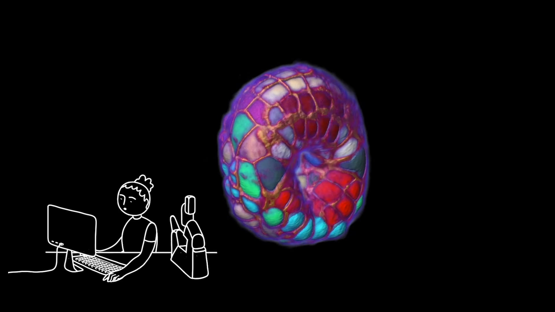
Building average representations of embryogenesis.

Our lab operates at the interface of computer science and developmental biology. We focus on the development of computational methods for analysing morphogenesis at the single-cell scale in whole organisms throughout their development.

Our primary objective is to apply computer vision, graph-based, machine learning and big data algorithms to quantify and comprehend developmental reproducibility during embryogenesis in various model organisms.

Our ultimate aim is to investigate the impact of developmental variability on morphogenesis by utilising homemade statistical averages from two main data modalities: fluorescence microscopy images and spatial omics.

Phallusia mammillata embryo

Media

Videos of developing embryos (usually)

Speaking

Guignard Lab

Channel info

Videos of embryonic development and related

Instructionals, Science & Technology, Documentary, Culture & Tech

embryo, developmental biology, Christophe Godin, Mouse Embryogenesis, Ulla Maj Fiuza, Development, Biology, cell biology, Single-Cell Resolution, Phallusia mammillata, animation, Philipp J. Keller, Ascidie, Fate Mapping, Howard Hughes Medical Institute, biology, Tracking, Light-Sheet Microscopy, Segmentation, cell signaling, Gregoire Malandain, Leo Guignard, Gastrulation, Early Organogenesis, science, Tissue Morphogenesis, ocean, Patrick Lemaire, Live Cell Imaging, Katie McDole

Guignard Lab

Cries or whispers between cells

Read more Like us, the cells of living beings communicate with each other to coordinate their behaviour. This short animated outreach video, directed by Laurence Serfaty and Isis Leterrier, explains how cells communicate during the embryonic development of animals. It is based on a scientific study published on July 10 2020 in the magazine "Science" by a team of researchers, biologists, computer scientists and physicists from CNRS, INRIA and EMBL: Guignard L, Fiúza UM, Leggio B, Laussu J, Faure E, Michelin G, Biasuz K, Hufnagel L, Malandain G, Godin C, Lemaire P. Contact area-dependent cell communication and the morphological invariance of ascidian embryogenesis. Science. 2020 Jul 10;369(6500):eaar5663.

Documentary, Culture & Tech, Instructionals

science, biology, embryo, ocean, animation, cell biology, cell signaling, developmental biology

Ascidian development

Read more Developing P. mammillata, cells are segmented and tracked using ASTEC

Science & Technology

Leo Guignard, Ulla Maj Fiuza, Patrick Lemaire, Christophe Godin, Development, Light-Sheet Microscopy, Tissue Morphogenesis, Single-Cell Resolution, Biology, Ascidie, Fate Mapping, Gastrulation, Gregoire Malandain, Live Cell Imaging, Phallusia mammillata, Segmentation, Tracking

10 Phallusia embryos | Fluo microscopy | Segmentation

Read more 10 Phallusia mammillata embryos. 3D volume rendering of fluorescent images and their segmentations and tracking. Colors show different tissues

Science & Technology

Leo Guignard, Christophe Godin, Patrick Lemaire, Gregoire Malandain, Development, Fate Mapping, Gastrulation, Light-Sheet Microscopy, Live Cell Imaging, Single-Cell Resolution, Tissue Morphogenesis, Ulla Maj Fiuza, Segmentation, Tracking, Ascidie, Phallusia mammillata, Biology

Dynamic In Toto Imaging of Post-Implantation Development / Cell Oct. 11, 2018 (Vol 175, Issue 3)

Read more McDole et al. develop a light-sheet microscopy approach and computational platform that accommodates the dynamics of post-implantation development to reconstruct at single-cell resolution mouse embryogenesis from gastrulation to early organogenesis. Check out the paper at: http://www.cell.com/cell/fulltext/S0092-8674(18)31243-1 Katie McDole, Léo Guignard, Fernando Amat, Andrew Berger, Grégoire Malandain, Loïc A. Royer, Srinivas C. Turaga, Kristin Branson, and Philipp J. Keller. In Toto Imaging and Reconstruction of Post-Implantation Mouse Development at the Single-Cell Level. Cell 175. And read more great research at http://www.cell.com/cell/home.

Science & Technology

Development, Mouse Embryogenesis, Gastrulation, Early Organogenesis, Tissue Morphogenesis, Live Cell Imaging, Fate Mapping, Single-Cell Resolution, Light-Sheet Microscopy, Katie McDole, Philipp J. Keller, Howard Hughes Medical Institute

15.07.2020 | Sea squirt development movie

Read more Des embryons d'ascidie (P. Mammillata) en cours de développement ont été imagés avec un microscope à feuille de lumière et traités informatiquement pour identifier et reconstruire chaque cellule. Les couleurs des reconstructions indiquent les tissus que chaque cellule formera (exemple : les cellules fuchsia formeront les muscles de la queue, les cellules vert foncé et cyan formeront le cerveau). © Léo Guignard Contact-area dependent cell communication and the morphological invariance of ascidian embryogenesis Guignard L, Fiuza UM, Leggio Laussu Faure E, Michelin G, Biasuz K, Hufnagel L, Malandain G, Godin C, Lemaire P Science, July 10 2020 , https://science.sciencemag.org/content/369/6500/eaar5663

Science & Technology

bottom of page